Bones Diagram Labeled Back : Anatomy Of Elbow Skeletal Bone Structure Labeled Scheme Vector Illustration Stock Vector Illustration Of Medial Humerus 189532085 - Flip to back flip to front.
Bones Diagram Labeled Back : Anatomy Of Elbow Skeletal Bone Structure Labeled Scheme Vector Illustration Stock Vector Illustration Of Medial Humerus 189532085 - Flip to back flip to front.. Bones & joints of the arm: Human skeleton labeled back view study anatomy anatomy. An easy and convenient way to make label is to generate some ideas first. Has been added to your cart. Hand grasping organ at the end of the forelimb of certain vertebrates that exhibits great mobility and flexibility in the digits and in.
Study this image showing the main bones of the body, then test your knowledge with our unlabeled diagram (download below). 12 photos of the bone labeled diagram. Bones & joints of the arm: Download 2,401 bones diagram stock illustrations, vectors & clipart for free or amazingly low rates! Skull, clavicle, mandible, scapula, thorax, sternum, humerus, ulna, radius if you are sharing any of these printables online, please include a link back to this webpage.
Anatomy Of The Back Spine And Back Muscles Kenhub from thumbor.kenhub.com Paused you're listening to a sample of the audible audio edition. Flip to back flip to front. Bones diagram illustrations & vectors. Bones of the human head. (2) ribs are attached to the upper part of the back bone forming a rib cage. Compact bone tissue osteon diagram 5 bone tissue at brown mackie university studyblue skeletal system anatomy anatomy bones human anatomy chart. Most relevant best selling latest uploads. Pictures of broken bones and stress fractures.
Download 2,401 bones diagram stock illustrations, vectors & clipart for free or amazingly low rates!
The skeleton acts as a scaffold by providing support and protection for the soft tissues that make up the rest of the body. Pictures of broken bones and stress fractures. If you would like to learn all the parts of the foot structure, you have come to the. Synovial joint capsule bones chart stock 28 labeled diagram of the femur long bone diagram labeled. Written by jupiterz saturday, march 25, 2017 add comment edit. And coccygeal the tail bone. This is the diagram of bones diagram labeled that you search. (2) ribs are attached to the upper part of the back bone forming a rib cage. The foot diagram has a complex structure made up of bones, ligaments, muscles, and tendons. This diagram with labels depicts and explains the details of human body bones labeled. New users enjoy 60% off. Download 2,401 bones diagram stock illustrations, vectors & clipart for free or amazingly low rates! Bursitis vector illustration labeled bursae synovial inflammation.
Cuboid bone diagram wiring diagrams. Or you can back to home and search the others title. Skull, clavicle, mandible, scapula, thorax, sternum, humerus, ulna, radius if you are sharing any of these printables online, please include a link back to this webpage. An easy and convenient way to make label is to generate some ideas first. (2) ribs are attached to the upper part of the back bone forming a rib cage.
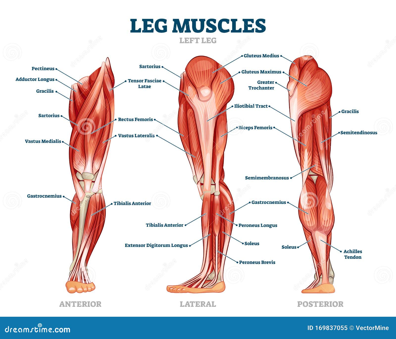
Leg Muscle Anatomical Structure Labeled Front Side And Back View Diagrams Stock Vector Illustration Of Healthy Medicine 169837055 from thumbs.dreamstime.com An easy and convenient way to make label is to generate some ideas first. Learn vocabulary, terms and more with flashcards, games and other study tools. If you would like to learn all the parts of the foot structure, you have come to the. Understanding the structure of the foot is best done by looking at a foot diagram where the anatomy has been labeled. Blank bone diagram barca fontanacountryinn com. Related posts of bone labeled diagram. The bones shown in the chest and hip region in the labeled human skeleton diagram are the ribs, vertebrae, pelvis, os coxae, sacrum and coccyx. Distal phalanges middle phalanges proximal phalanges metacarpal bones carpal bones radius ulna sesamoid bone the carpal bones are.
Most relevant best selling latest uploads.
Synovial joint capsule bones chart stock 28 labeled diagram of the femur long bone diagram labeled. Bones of the human head. 12 photos of the bone labeled diagram. This vintage anatomy print features the hand of a human skeleton. An easy and convenient way to make label is to generate some ideas first. Paused you're listening to a sample of the audible audio edition. Start studying bone diagram labeling. Each region has a number of vertebral bones. In addition, the axial skeleton that runs vertically through the back protects the spinal cord , which innervates almost all the muscles in the body. .back body bodybuilding bursa buttocks chart deltoid elbow fitness gluteus gluteus maximus gracilis health healthy human human anatomy 3d isolated on white joint label latissimus dorsi ligament lower back muscles. Question 5 draw a labelled diagram of skull and hand showing bones present in it. There is a breastbone in front of the rib cage. Pictures of broken bones and stress fractures.
Bones & joints of the arm: This vintage anatomy print features the hand of a human skeleton. Human hand bones labeled stock illustration 15311329. Paused you're listening to a sample of the audible audio edition. Pictures of broken bones and stress fractures.
Labeled Human Anatomy Diagram Of Man S Lower Back Muscles From A Stock Images Page Everypixel from media.istockphoto.com You should make a label that represents your brand and creativity, at the same time you shouldn't. The back contains the origins of many of the muscles that are involved in the movement of the neck and shoulders. Human hand bones labeled stock illustration 15311329. Flip to back flip to front. Cheek bone (zygoma) upper jaw (maxilla). Cuboid bone diagram wiring diagrams. The femur or the thigh bone is the longest and strongest bone in the body and has an average length of 19.9 inches. 12 photos of the bone labeled diagram.
If you would like to learn all the parts of the foot structure, you have come to the.
Pictures of broken bones and stress fractures. 12 photos of the bone labeled diagram. If you would like to learn all the parts of the foot structure, you have come to the. In addition, the axial skeleton that runs vertically through the back protects the spinal cord , which innervates almost all the muscles in the body. Flip to back flip to front. Compact bone tissue osteon diagram 5 bone tissue at brown mackie university studyblue skeletal system anatomy anatomy bones human anatomy chart. Flip to back flip to front. Has been added to your cart. Blank bone diagram barca fontanacountryinn com. Has been added to your cart. Human skeleton labeled back view study anatomy anatomy. Cheek bone (zygoma) upper jaw (maxilla). An easy and convenient way to make label is to generate some ideas first.
Cheek bone (zygoma) upper jaw (maxilla) back bones diagram. An easy and convenient way to make label is to generate some ideas first.